


Medical scans and diagnostic images often vary in appearance from one hospital, scanner, or dataset to another, even when they capture the exact same organ or disease. Because standard deep learning models rely heavily on image intensity and surface appearance, a system trained on scans from one clinic frequently makes errors when analysing images from a different source. To overcome this drop in cross-dataset accuracy, researchers at SRM AP developed DGSA-Net, a structural attention network that prioritises the underlying shape, edges, and geometric layout of anatomical regions rather than raw pixel brightness. By learning what a structure looks like regardless of variations in scan appearance, the framework produces more consistent segmentation of lesions, tumours, and organs across different medical imaging datasets.
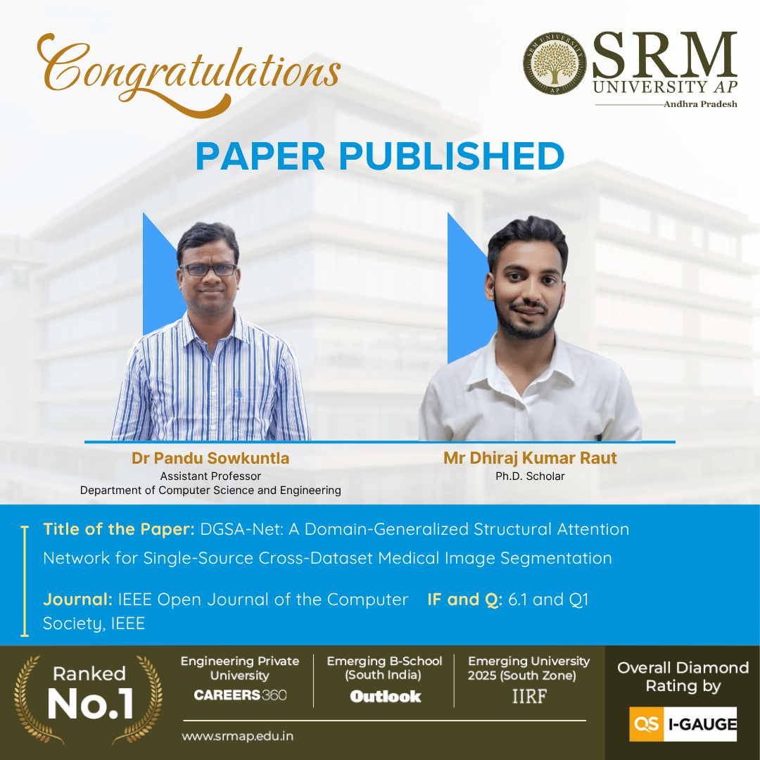
In a study published in IEEE Open Journal of the Computer Society, a Q1 journal with an impact factor of 6.1 published by IEEE, Dr Pandu Sowkuntla, Assistant Professor in the Department of Computer Science and Engineering at SRM University-AP, led this work alongside PhD Scholar Mr Dhiraj Kumar Raut. Their published paper is titled “DGSA-Net: A Domain-Generalized Structural Attention Network for Single-Source Cross-Dataset Medical Image Segmentation“.
Medical image segmentation models often perform well on the datasets on which they are trained, but their performance may degrade when they are applied to images collected under different acquisition conditions or from different institutions. This work proposes DGSA-Net, a structural attention-based deep learning framework designed to improve robustness under such cross-dataset domain shifts. The model combines a Structural Attention Refinement Module (SARM), hierarchical cross-scale feature fusion, boundary-aware progressive decoding, and a structure-constrained hybrid loss. The framework was evaluated on dermoscopy, CT, and chest X-ray datasets under both in-domain and unseen cross-dataset settings. The results show improved segmentation accuracy, boundary preservation, and reduced generalization gap compared with several established CNN-, Transformer-, foundation-model-, and domain-generalization-based approaches.
The framework supports computer-aided medical image analysis when a segmentation model trained at one facility must process scans acquired from another hospital, imaging device, or clinical setting. By tolerating differences in image acquisition and annotation protocols, DGSA-Net can reduce the need for repeated dataset-specific retraining while helping clinicians outline lesions and anatomical structures for quantitative assessment, treatment planning, and disease monitoring. Because the system is currently at the research stage, prospective and multicentre clinical validation is needed before hospital deployment. Next, Dr Pandu Sowkuntla and Mr Dhiraj Kumar Raut plan to test DGSA-Net across more varied source-target dataset pairs, extend the architecture from 2D binary tasks to 3D and multi-class medical image segmentation, evaluate alternative encoders beyond ResNet-34, and examine how dataset-specific preprocessing and annotation standards influence model performance.
Read the full article here