
 Alzheimer’s disease slowly damages the brain and affects memory, thinking, and daily activities. Detecting it early can help doctors begin treatment sooner. Dr Sarvani Anandarao, Assistant Professor in the Department of Computer Science and Engineering at SRM AP, in her research titled “Differential-Evolution-Optimized Swin Transformer–Temporal Convolutional Network for Explainable MRI-Based Alzheimer’s Disease Classification“published on the Q2 journal of Ingénierie des Systèmes d’Information (ISI) having an impact factor of 1.74, has developed an Artificial Intelligence (AI) system that studies MRI brain scans and automatically determines whether a person is healthy or has Alzheimer’s disease.
This research was carried out through collaboration among researchers from
- RBC Wealth Management, Minneapolis, USA
- Publicis Sapient, Minneapolis, USA
- VIT-AP University, India, and
- SRM University-AP, India
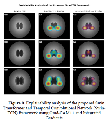
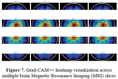
Unlike many existing AI systems, this model also explains why it made a particular decision by highlighting the affected areas of the brain in the MRI image. This makes the technology easier for doctors to trust and use in real clinical practice. This AI system achieved 99.5% accuracy, showing that it can assist healthcare professionals in making faster and more reliable diagnoses.
Abstract
Alzheimer’s disease (AD) is a progressive neurological disorder in which early diagnosis is essential for timely intervention. This research proposes an explainable deep learning framework that combines a Swin Transformer for hierarchical spatial feature extraction, a Temporal Convolutional Network (TCN) for modeling inter-slice relationships, and a Hybrid Attention Mechanism (HAM) to emphasise diagnostically relevant MRI features. Differential Evolution (DE) is employed to optimise key hyperparameters, improving model performance and stability. The proposed framework was evaluated on a publicly available MRI Alzheimer’s dataset using subject-wise five-fold cross-validation and achieved 99.50% classification accuracy with an AUC of 0.92. Explainable AI techniques, including Grad-CAM++ and Integrated Gradients, were used to visualise the brain regions influencing model decisions, making the framework more transparent and clinically interpretable.
Practical Applications and social Implications
The proposed research has several important practical applications, starting with the early detection of Alzheimer’s disease before severe cognitive decline occurs. By serving as a decision-support tool for neurologists and radiologists, this technology reduces manual MRI interpretation time and improves diagnostic consistency by minimising human error. Furthermore, its use of explainable AI enables clinicians to understand and trust the AI predictions, allowing for potential integration into hospital Computer-Aided Diagnosis (CAD) systems. Ultimately, this research supports better treatment planning and patient monitoring, which can contribute to reducing healthcare costs associated with delayed diagnosis and may improve the quality of life for patients and their families through earlier intervention.
Future work will focus on validation using larger multi-center clinical MRI datasets, which is consistent with the paper’s discussion that further validation on external clinical datasets is needed before clinical deployment. Additionally, future efforts will target the integration of multi-modal medical data such as PET scans, CT scans, and clinical information, alongside the early prediction of Mild Cognitive Impairment (MCI) progression. The research will also aim at the development of lightweight AI models suitable for deployment in hospitals, the creation of real-time clinical decision-support systems, and the utilisation of federated learning for privacy-preserving medical AI. Finally, future directions include extending the framework to detect other neurological disorders such as Parkinson’s disease and brain tumors, as well as incorporating enhanced explainability techniques to further improve clinician confidence.